This manual provides a comprehensive‚ hands-on approach to understanding human anatomy and physiology‚ offering 27 full-color exercises and a built-in Histology Atlas for interactive learning.
1.1 Purpose and Structure of the Manual
The manual is designed for a one-semester A&P course‚ offering 27 hands-on exercises and a Histology Atlas. It includes pre-lab quizzes‚ learning objectives‚ and review sheets‚ structured to complement Elaine Marieb’s textbook. The manual provides a clear‚ interactive approach for health-related programs‚ ensuring students gain practical skills and foundational knowledge in anatomy and physiology.
1.2 Key Features and Benefits
This manual offers 27 full-color‚ hands-on exercises‚ a built-in Histology Atlas‚ and pre-lab quizzes to enhance learning. It includes review sheets‚ learning objectives‚ and checklists of materials for organized study. The structured approach simplifies complex topics‚ ensuring students grasp essential concepts and apply them effectively in a fast-paced‚ one-semester A&P course.
Anatomical Language and Orientation
This section introduces foundational terminology and spatial concepts‚ enabling students to describe body structures accurately and understand directional terms essential for studying anatomy and physiology.
2.1 Key Terms and Definitions
This section introduces foundational anatomical terminology‚ including terms like ventral‚ dorsal‚ sagittal‚ and transverse planes‚ as well as proximal and distal. These definitions are crucial for accurately describing body structures and their spatial relationships‚ enabling students to communicate effectively in scientific and clinical settings. Understanding these terms is essential for mastering the language of anatomy and physiology.
2.2 Body Planes and Directions
The three main body planes—sagittal‚ frontal (coronal)‚ and transverse (horizontal)—divide the body into sections. Directions like anterior‚ posterior‚ superior‚ inferior‚ proximal‚ and distal help locate structures. These planes and terms are essential for describing movements and spatial relationships‚ enabling precise communication in anatomical and physiological studies. Mastery of these concepts aids in visualizing and understanding body organization and function.
2.3 Anatomical Regions and Cavities
Anatomical regions divide the body into manageable areas for study‚ such as the abdominal‚ thoracic‚ and pelvic regions. Cavities‚ like the cranial and thoracic‚ house vital organs. These sections help in locating structures and understanding internal organization. The manual emphasizes identifying and mapping these regions and cavities‚ crucial for medical terminology and diagnostics‚ enhancing comprehension of human anatomy and its functional layout.

The Human Body and Its Organization
This section explores the human body’s structural hierarchy‚ from cells to organ systems‚ emphasizing how these components function together to maintain overall health and bodily processes.
3.1 Levels of Organization
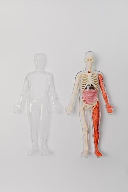
The human body is organized into a hierarchy of structural levels‚ beginning with cells‚ which form tissues‚ then organs‚ and finally organ systems. These levels work together to maintain homeostasis and overall function. Understanding this organization is crucial for comprehending how the body operates as an integrated whole‚ from basic cellular functions to complex systemic processes.
3.2 Organ Systems and Their Functions
Organ systems‚ such as the skeletal‚ muscular‚ nervous‚ and circulatory systems‚ work together to maintain homeostasis. Each system has distinct functions: the skeletal system provides support‚ the muscular system enables movement‚ the nervous system controls body activities‚ and the circulatory system transports essential nutrients and oxygen. Understanding their interdependence is key to grasping human physiology and overall bodily functions.
The Cell: Basic Unit of Life
The cell is the fundamental unit of life‚ performing essential functions like metabolism‚ reproduction‚ and DNA storage. Its structure‚ including the cell membrane‚ cytoplasm‚ and organelles‚ enables these processes.
4.1 Cell Structure and Components
The cell is the basic structural and functional unit of life‚ consisting of a cell membrane‚ cytoplasm‚ and organelles like the nucleus‚ mitochondria‚ ribosomes‚ and endoplasmic reticulum. These components work together to regulate cell growth‚ energy production‚ and reproduction‚ ensuring proper cellular function and maintenance. Understanding cell structure is essential for studying human anatomy and physiology‚ forming the foundation of life processes.
4.2 Cell Transport Mechanisms
Cells use various transport mechanisms to move materials across membranes‚ including passive transport (diffusion‚ osmosis) and active transport. Passive transport relies on concentration gradients‚ while active transport requires energy. Vesicular transport involves membrane vesicles for larger molecules. These mechanisms are crucial for maintaining homeostasis‚ enabling cells to exchange nutrients‚ waste‚ and signaling molecules efficiently‚ ensuring proper cellular function and overall health.
4.3 Cell Division and Growth
Cell division and growth are essential processes for tissue repair‚ development‚ and reproduction. Mitosis ensures genetic continuity by dividing cells into identical daughter cells‚ while cytokinesis completes cell division. Growth involves increased cell size and protein synthesis. Proper regulation of these processes is critical for maintaining health‚ preventing abnormal cell growth‚ and ensuring the body’s functional integrity throughout life.
Basic Tissues and the Integumentary System
This section explores the four primary tissue types—epithelial‚ connective‚ muscle‚ and nervous—and their functions. It also examines the integumentary system‚ focusing on skin structure and its protective roles in maintaining bodily homeostasis.
5.1 Types of Tissues
This section introduces the four primary types of tissues: epithelial‚ connective‚ muscle‚ and nervous. Each tissue is defined by its structure and function‚ with epithelial forming linings‚ connective supporting and connecting organs‚ muscle enabling movement‚ and nervous transmitting signals. Laboratory exercises provide hands-on identification of tissue samples‚ reinforcing their roles in maintaining bodily functions and overall health.
5.2 Skin Structure and Functions
The skin‚ the body’s largest organ‚ consists of the epidermis‚ dermis‚ and hypodermis. It protects against external damage‚ regulates temperature‚ and aids in sensation and secretion. Laboratory exercises explore skin layers‚ sweat and sebaceous glands‚ and the role of skin in maintaining homeostasis. Interactive activities highlight the skin’s essential functions‚ reinforcing its importance in overall health and bodily protection.

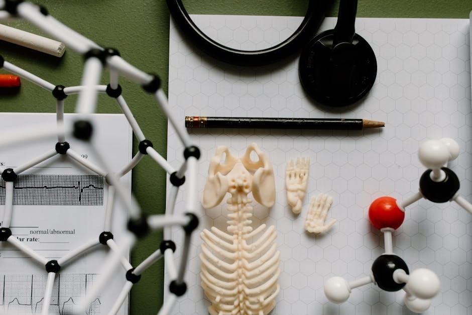
The Skeletal System
The skeletal system comprises bones and joints‚ providing structural support‚ protection‚ and movement. It also produces blood cells and stores minerals‚ essential for bodily functions.
6.1 Bones and Cartilage
Bones and cartilage form the skeletal system’s foundation. Bones provide structural support‚ protect organs‚ and facilitate movement. They also produce blood cells and store minerals like calcium. Cartilage‚ found in joints and between vertebrae‚ cushions and reduces friction. Both tissues are essential for mobility and overall bodily functions.
6.2 Joints and Movement
Joints‚ or articulations‚ are points where bones connect‚ enabling movement. They are classified as synovial‚ cartilaginous‚ or fibrous‚ with synovial joints allowing the greatest mobility. Ligaments and tendons support joints‚ facilitating actions like walking or running. Understanding joint structure and function is crucial for analyzing movement and the skeletal system’s role in locomotion.
The Muscular System
The muscular system consists of skeletal‚ smooth‚ and cardiac muscles‚ enabling movement‚ support‚ and posture maintenance. It functions with the nervous system to coordinate body activities effectively.
7.1 Types of Muscles
The muscular system comprises three types: skeletal‚ smooth‚ and cardiac muscles. Skeletal muscles are voluntary‚ attached to bones‚ enabling movement and posture; Smooth muscles are involuntary‚ found in internal organs‚ facilitating functions like digestion. Cardiac muscle is specialized for the heart‚ ensuring rhythmic contractions. Each type varies in structure‚ function‚ and control mechanisms‚ working collectively with the nervous system to maintain body activities and overall physiological balance effectively.
7.2 Muscle Structure and Contraction
Muscle contraction occurs due to the sliding filament theory‚ where actin and myosin filaments slide past each other‚ shortening the sarcomere. This process is regulated by neurotransmitters from motor neurons‚ triggering calcium release and contraction. The sarcomere‚ the functional unit of muscle‚ consists of Z-lines‚ A-bands‚ and I-bands. ATP hydrolysis provides energy‚ enabling cross-bridge formation and relaxation‚ allowing muscles to move bones and maintain posture effectively.
The Nervous System
The nervous system consists of the central and peripheral nervous systems‚ functioning to transmit and process information; Neurons communicate via synapses‚ enabling regulation of body functions and responses to stimuli.
8.1 Neurons and Neuroglia
Neurons are specialized cells that transmit nerve impulses‚ consisting of dendrites‚ a cell body‚ and an axon. Neuroglia‚ such as astrocytes and Schwann cells‚ support neurons by providing nutrients‚ removing waste‚ and forming myelin sheaths to enhance nerve signal transmission‚ ensuring efficient communication within the nervous system.
8.2 Reflexes and Nerve Impulses
Reflexes are automatic responses to stimuli‚ involving a reflex arc with sensory receptors‚ afferent neurons‚ the central nervous system‚ efferent neurons‚ and effectors. Nerve impulses‚ or action potentials‚ are generated by ion exchanges across cell membranes‚ transmitting signals through neurons. Synapses‚ where neurons communicate via neurotransmitters‚ enable complex functions like movement and thought‚ forming the basis of the nervous system’s functionality.

The Circulatory System
The circulatory system‚ including blood‚ blood vessels‚ and the heart‚ is explored through hands-on exercises‚ providing a detailed understanding of its structure and essential functions.
9.1 Blood and Blood Vessels
Blood is a liquid tissue consisting of plasma‚ red blood cells‚ white blood cells‚ and platelets‚ essential for oxygen transport‚ immune defense‚ and clotting. Blood vessels‚ including arteries‚ veins‚ and capillaries‚ form a closed system for circulating blood throughout the body. Arteries carry oxygen-rich blood away from the heart‚ while veins return oxygen-poor blood. Capillaries enable nutrient and waste exchange at the cellular level‚ maintaining homeostasis and overall health.
9.2 Heart Structure and Function
The heart is a muscular organ with four chambers: the right and left atria‚ and right and left ventricles. The septum separates the chambers‚ while valves ensure one-way blood flow. The heart receives deoxygenated blood through the right atrium‚ pumps it to the lungs‚ and returns oxygen-rich blood to the left side for distribution. This dual circulation maintains oxygen supply and overall bodily function‚ essential for survival.
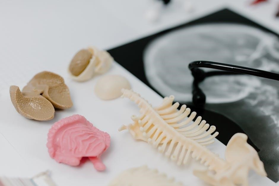
Histology and Microscopic Anatomy
Histology explores the microscopic structure of tissues‚ using techniques like sectioning and staining to examine cell arrangements. This field is crucial for understanding tissue function and pathology.
10.1 The Compound Light Microscope
The compound light microscope is a fundamental tool in histology‚ enabling detailed examination of tissue structures. It uses a combination of objective and eyepiece lenses to magnify specimens. Proper preparation of slides‚ including sectioning and staining‚ is essential for clear observation. The microscope’s illumination system enhances visibility‚ making it indispensable for studying cellular details in anatomy and physiology laboratories. Its versatility aids both students and researchers in exploring microscopic anatomy effectively.
10.2 Preparing and Examining Tissue Samples
Preparing tissue samples involves sectioning‚ staining‚ and mounting to ensure proper visualization; Techniques like fixation preserve tissue integrity‚ while staining enhances contrast for microscopic examination. Proper preparation is critical for accurate histological analysis. This process allows students to observe cellular structures and understand tissue organization‚ integrating with compound light microscopy for a comprehensive study of microscopic anatomy and physiology.
Laboratory Exercises and Investigations
This section provides 27 hands-on exercises‚ including pre-lab quizzes‚ safety protocols‚ and review sheets‚ to enhance practical understanding of human anatomy and physiology.
11.1 Pre-Lab Preparation and Safety
This section outlines essential steps for preparing for laboratory exercises‚ including material checklists‚ safety protocols‚ and pre-lab quizzes to ensure a safe and effective learning environment.
11.2 Conducting Hands-On Activities
This section focuses on engaging‚ interactive exercises that allow students to explore anatomical structures and physiological processes firsthand. Activities include dissections‚ histology slides‚ and physiological measurements‚ supported by clear instructions and visual aids to enhance understanding. Students are encouraged to work in groups‚ fostering collaboration and practical application of concepts learned in the textbook.
11.3 Histology and Dissection Labs
These labs provide hands-on experience with histological slides and dissection of specimens‚ enabling students to examine microscopic structures and organ systems in detail. Activities include identifying tissue types under a microscope and exploring anatomical relationships through dissection. The exercises are supported by visual guides and safety protocols to ensure a safe and effective learning environment.
Review and Assessment Tools
The manual includes quizzes‚ review sheets‚ and lab reports to reinforce learning and assess understanding‚ providing students with clear tools to track their progress effectively.
12.1 Quizzes and Review Sheets
The manual features quizzes and review sheets to assess understanding and retention of key concepts. These tools include multiple-choice questions‚ fill-in-the-blank exercises‚ and labeling activities. Designed to complement lab exercises‚ they help students identify areas for further study and ensure mastery of essential anatomy and physiology principles before progressing to more complex topics.
12.2 Laboratory Reports and Presentations
Laboratory reports and presentations are integral to the learning process‚ allowing students to document and communicate their findings effectively. The manual provides structured formats for reports‚ including sections for introduction‚ methods‚ results‚ and conclusions. Students are encouraged to use visual aids like graphs and images to enhance their presentations‚ ensuring clear and concise communication of scientific concepts and data analysis.
Integration with Other A&P Resources
The manual is designed to complement textbooks and digital tools‚ offering interactive simulations‚ quizzes‚ and multimedia content to enhance student engagement and understanding of A&P concepts.
13.1 Companion Textbooks
The manual is most effectively paired with Marieb’s Essentials of Human Anatomy and Physiology textbooks‚ such as the 12th or 14th editions‚ offering a seamless learning experience. It complements the core content with hands-on exercises‚ ensuring a comprehensive understanding of A&P concepts through interactive and visual learning tools;
13.2 Digital and Online Supplements
The manual is supported by digital and online resources‚ including eTextbooks and interactive platforms‚ offering flexible learning options. Students can access virtual labs‚ quizzes‚ and multimedia content to enhance their understanding. These supplements provide self-assessment tools‚ video tutorials‚ and 3D models‚ ensuring a dynamic and engaging learning experience that complements the lab manual’s hands-on activities.
This manual equips students with practical skills in anatomy and physiology‚ preparing them for real-world applications in healthcare and encouraging continuous learning in the field.
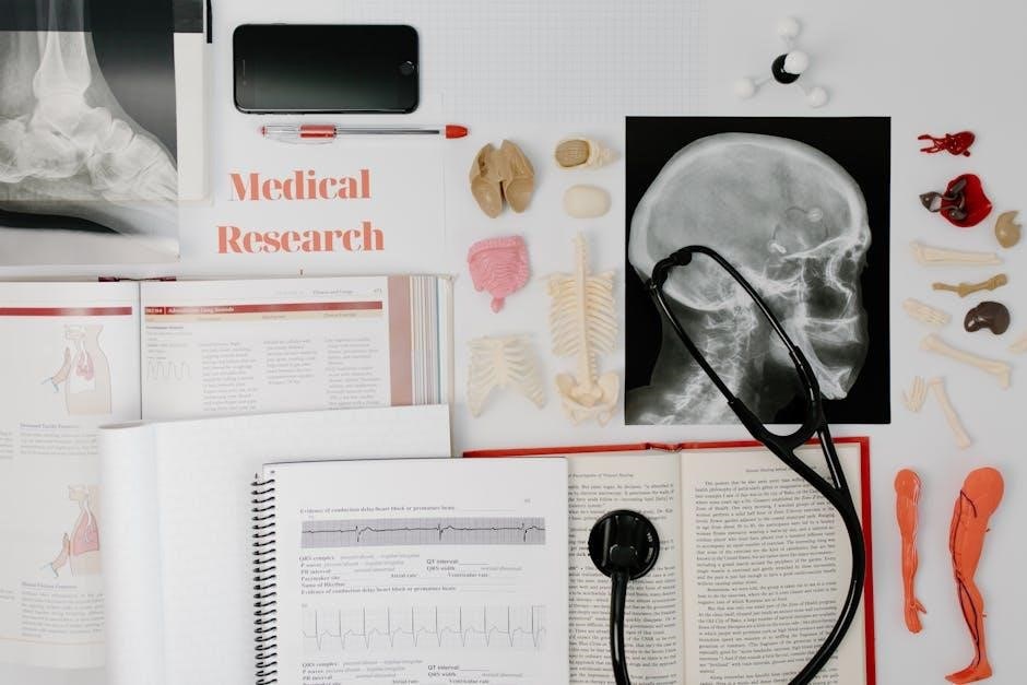
14.1 Applying Lab Skills in Healthcare
The manual’s hands-on exercises prepare students for real-world healthcare applications‚ enabling them to apply anatomical and physiological knowledge in clinical settings. Lab skills such as histology‚ dissection‚ and data analysis are vital for careers in medicine‚ nursing‚ and research. Understanding human structure and function is essential for accurate diagnostics‚ treatments‚ and patient care‚ making this manual a valuable tool for future healthcare professionals.
14.2 Continuous Learning in A&P
Continuous learning in anatomy and physiology is essential for healthcare professionals‚ as advancements in medical science evolve rapidly. This manual supports lifelong learning by providing interactive exercises‚ review sheets‚ and a histology atlas‚ enabling students to reinforce concepts and develop practical skills. Its structured approach ensures a strong foundation‚ preparing learners for ongoing education and professional challenges in the dynamic field of healthcare.